
English 


Author: Site Editor Publish Time: 2026-10-04 Origin: Site


High-quality intraoral imaging is no longer just a simple diagnostic aid. It serves as the primary driver for patient case acceptance in modern dentistry. However, the market remains heavily saturated. Manufacturers constantly push conflicting resolution claims. Procurement managers and clinical directors face a difficult challenge. They often overpay for misleading marketing metrics, like inflated megapixels. They frequently overlook the underlying sensor technology. They ignore critical software bottlenecks and everyday clinical usability.
You need a reliable way to cut through this industry noise. Poor image quality directly impacts case presentation. It damages patient trust. This guide provides an objective, evidence-based framework to evaluate intraoral camera resolution. We will deeply compare standard and emerging formats. You will understand the massive differences between legacy systems and modern marvels. You will learn how to de-risk the entire procurement process. We will help you select the exact equipment your practice actually needs to succeed.
720P is obsolete for modern diagnostic standards but remains in legacy systems; 1080P is the current baseline for standard clinical workflows; 6K offers future-proof macro detailing but requires upgraded display infrastructure.
Resolution is only as valuable as the camera's optical capability; a high resolution dental camera must be paired with superior lighting and optics.
Evaluating a dental camera supplier requires looking beyond spec sheets to assess software compatibility, warranty terms, and true sensor size.
Adoption success hinges on network bandwidth and operatory monitor limitations—deploying a 6K intraoral camera on a 1080P operatory monitor yields zero visible ROI.
Clinicians utilize intraoral cameras for two distinct purposes. They need absolute diagnostic confidence. They also require highly effective patient communication tools. You must clarify this dual purpose before purchasing new equipment. Older 720P sensors might help a dentist spot a gross fracture. However, modern dentistry demands much more precision. Higher resolutions become strictly necessary for complex cases. You need superior pixel density to show patients micro-leakage. You need it to highlight tiny hairline cracks. Operatory screens now frequently exceed 40 inches in size. Displaying a low-resolution image on a massive screen causes severe pixelation. The image becomes a blurry grid. Patients cannot understand what they are looking at.
Consider your daily case acceptance metrics. Crystal-clear imagery correlates directly to higher patient trust. Patients rarely accept expensive treatment plans based on verbal descriptions alone. They need visual proof. High-definition visuals remove all ambiguity from the conversation. When patients clearly see failing margins, they agree to treatment faster. You spend less time convincing them. You spend more time performing dentistry. Clear imagery acts as your best case presentation tool.
Insurance claim approvals represent another massive financial factor. Dental insurance payers constantly demand undeniable proof of medical necessity. Blurry or artifact-heavy images lead directly to claim denials. Artifact-free, crisp images reduce these frustrating rejections. Clear visual evidence accelerates your reimbursement cycles significantly. You protect your practice revenue stream. You reduce the administrative burden on your front office team. High-resolution imagery easily pays for itself through increased approvals.
We must evaluate different resolution tiers objectively. Each tier serves a very specific clinical function. Practice owners must align these capabilities against their actual daily procedures. Buying too little resolution hurts diagnostics. Buying too much resolution strains your IT infrastructure.
Resolution Tier | Primary Use Case | Key Benefits | Major Limitations |
|---|---|---|---|
720P (HD) | Basic visual aids, older practice IT setups | Low bandwidth, cheap replacement cost | Severe pixelation on large monitors, poor detail |
1080P (FHD) | General restorative and daily hygiene | Universal compatibility, perfect file size balance | Lacks macro-level zooming capabilities |
6K (Ultra HD) | Cosmetic dentistry, endodontics, premium clinics | Unmatched micro-detail, lossless digital zoom | Requires massive storage and 4K/6K monitors |
The 720P format represents the legacy budget tier. It serves merely as a basic visual aid today. It fits older existing practice infrastructure perfectly. Legacy USB 2.0 connections handle its small data load easily. However, it carries significant clinical limitations. You experience heavy pixelation when scaling images on modern monitors. The small sensor lacks the necessary detail. It cannot capture fine marginal discrepancies accurately. You should actively phase out this resolution.
The 1080P format stands as the current clinical standard. Most practices rely entirely on this resolution daily. It acts as the reliable workhorse for 80% of daily restorative workflows. Hygienists use it constantly for patient education. It offers excellent operational benefits. You get a perfect balance of image clarity and manageable file sizes. It also ensures universal software compatibility. Almost every practice management system handles 1080P images flawlessly. It provides the safest upgrade path.
The 6K format represents the emerging macro standard. This tier pushes digital imaging boundaries. It suits high-end cosmetic dentistry perfectly. Endodontists use it for intense macro-photography. It future-proofs premium practices against rapid technological shifts. You gain unmatched detail for identifying micro-fractures. It allows aggressive digital zooming without losing structural clarity. Investing in a true 6K intraoral camera transforms case presentations.
However, you must maintain a skeptical lens regarding 6K claims. Manufacturers frequently manipulate marketing materials. Beware of interpolated or upscaled 6K devices. Interpolation uses software algorithms to guess missing pixels. It creates artificial resolution. True 6K resolution requires a physically larger image sensor. It needs better glass optics. It cannot rely purely on cheap software enhancements.
Resolution numbers mean absolutely nothing without proper hardware mechanics backing them up. A sensor can boast millions of pixels. Yet, it will produce terrible images if the supporting technology fails. We must explore the underlying physical components.
Megapixels do not tell the whole diagnostic story. Sensor physical size matters immensely. Packing more pixels onto a tiny sensor surface causes severe image noise. The individual pixels become too small. They fail to capture enough light photons. This creates a grainy, unusable image. Larger physical sensors remain absolutely critical for true high-definition imaging. Modern CMOS sensors handle light gathering much better than older CCD models. They deliver crisper images inside the dark oral cavity. They process visual data faster.
Fixed focus lenses often ruin high-resolution sensors completely. A massive 6K sensor proves totally useless if the focal point stays blurry. You cannot diagnose a blurry margin. This reality makes an adjustable focus intraoral camera mandatory for serious clinical work. You need reliable liquid lens autofocus technology. Liquid lenses change shape instantly via electrical currents. They have no mechanical moving parts. They ensure sharp captures across vastly different depths of field. Mechanical voice coil autofocus is another viable option. It physically moves the lens element quickly. Both mechanisms guarantee optical sharpness before the image sensor captures the frame.
Lighting geometry makes or breaks your clinical image. The oral cavity presents a highly reflective environment. Wet enamel acts like a mirror. Built-in LED array design prevents harsh glare on these wet surfaces. Specular reflection easily ruins high-resolution captures. It creates massive white blow-out spots on the tooth. Proper illumination geometry diffuses LED light evenly. Some advanced designs incorporate cross-polarized filters. These filters cut through saliva glare entirely. They reveal the true underlying dentin shade accurately. They eliminate harsh reflections and deep shadows.
Deploying new imaging hardware introduces very unique operational challenges. You cannot simply plug a premium camera into an outdated computer. You must prepare your IT infrastructure beforehand. Failure to prepare leads to massive frustration.
First, consider the strict hardware bottleneck. Displaying an ultra-high-definition image demands compatible viewing hardware. You must use a fully compatible operatory monitor. A 1080P screen physically cannot display a 6K image. The operating system forcefully downsamples the image. It compresses the visual data to fit the smaller screen grid. You immediately lose all the extra detail you paid for. Upgrading your cameras often forces a simultaneous monitor upgrade across all operatories.
Second, evaluate your storage capacity and network bandwidth. High-resolution images generate massive raw file sizes. A single uncompressed frame can exceed 15 megabytes. This reality severely impacts your daily digital workflow. Evaluate the sudden impact on your cloud storage costs. Large files also slow down practice management software loading times. When a hygienist opens a patient chart, the software must download those huge files. A slow local network will cause the software to freeze. You might need Gigabit ethernet switches to handle the increased data load smoothly.
Third, address ergonomics and workflow friction. Larger image sensors require vastly different internal board designs. They often demand more internal cooling components. This complexity leads to noticeably heavier wands. Address operator fatigue early in the procurement phase. Hygienists hold these devices for extended periods daily. A heavy wand causes rapid wrist strain. Prioritize lightweight, highly ergonomic designs. Comfortable tools guarantee higher adoption rates among your clinical staff. A camera left in the drawer yields zero return on investment.
Selecting the right hardware partner protects your substantial capital investment. You need more than just impressive technical specifications. You need reliability, integration, and absolute transparency from your chosen vendor.
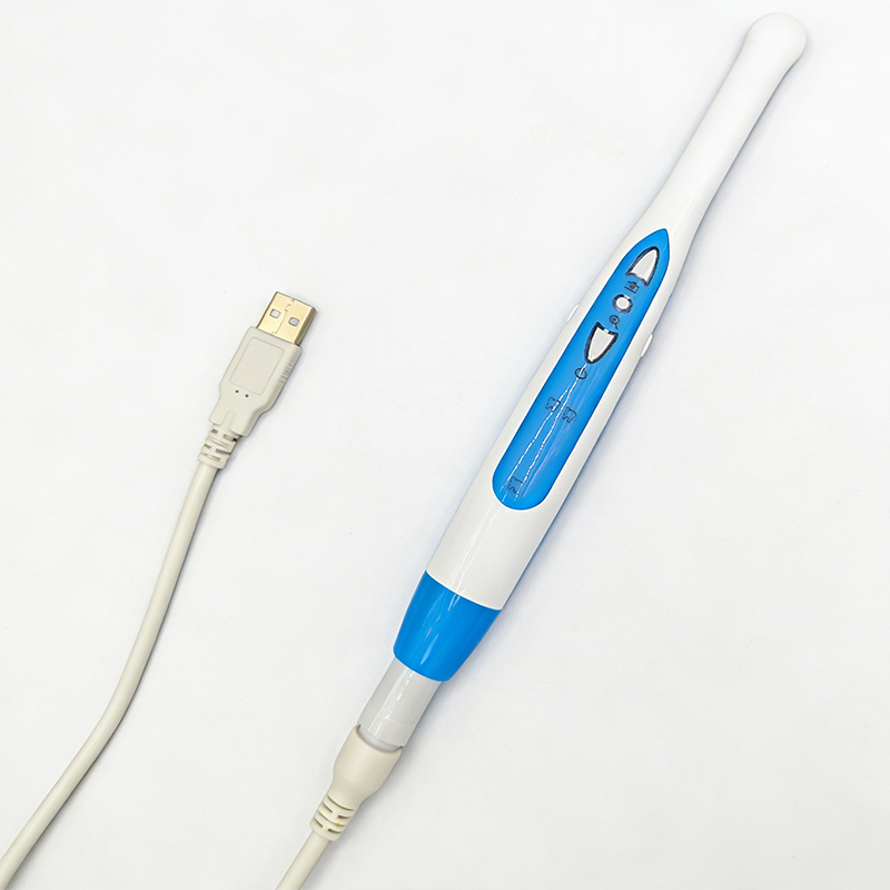
Proprietary software lock-ins create massive future headaches. Some manufacturers force you to use their specific viewing software. This fragments your workflow. The ideal USB intraoral camera manufacturer provides fully software-agnostic devices. They engineer cameras to natively integrate with major imaging software. This includes industry giants like Dexis, Eaglesoft, or Romexis. They achieve this using standard TWAIN protocols or direct API integrations. You avoid clunky third-party bridging software. Direct integration ensures the physical capture button on the wand works instantly. It maintains strict infection control protocols. Staff do not have to touch the computer mouse.
You must vet your potential providers carefully. When evaluating an OEM dental imaging camera provider, look far beyond their glossy brochure. Dig into their actual manufacturing standards. Check for strict ISO 13485 compliance in their medical device manufacturing process. This certification guarantees robust quality control. Demand clear supply chain transparency regarding their internal components. Investigate their return merchandise authorization rates. High return rates indicate severe factory defects. A truly reputable dental camera supplier will readily provide these reliability metrics upon request. They stand behind their engineering. They offer rapid hot-swap warranty programs. They ship loaner devices overnight to prevent clinical downtime.
You must actively align your hardware choices with your specific clinical needs. Buying blindly wastes capital. Follow this simple shortlisting logic. The 1080P tier remains the safest choice for general practices. It provides the most frictionless upgrade path from older legacy systems. It balances quality against IT demands perfectly. Alternatively, the 6K tier is fully justified for specialized cosmetic or endodontic practices. Just ensure you possess the robust IT infrastructure necessary to support it. Upgrading monitors and network speeds is mandatory for this tier.
A truly high resolution dental camera dramatically elevates your entire practice standard. It boosts case acceptance instantly. Take these concrete next steps today. Audit your current operatory monitors for native resolution capabilities. Check your current imaging software for TWAIN compatibility. Verify your local network upload speeds. Only issue a request for proposal after confirming your infrastructure readiness. Request a live clinical demo inside your own operatory. Test the wand weight in your own hands.
A: No. Resolution alone cannot overcome poor hardware design. Lighting geometry, physical sensor size, and optical focus mechanics remain equally important. Without a high-quality glass lens and proper LED illumination, extra pixels only capture blurry, unusable details.
A: It can. If your network infrastructure and storage servers lack optimization, large raw image files cause severe lag. You must ensure your local network bandwidth and cloud storage capacity can easily handle the increased daily data load.
A: USB models offer tremendous plug-and-play versatility. They work easily across completely different operatory computers. Proprietary connections often lock the practice into one specific software ecosystem. They restrict operational flexibility and make future technology upgrades far more difficult.
A: Check the native hardware resolution of the internal CMOS sensor. Ignore the "software enhanced" or "interpolated" maximum output numbers listed in marketing materials. Native resolution determines actual hardware capability. Interpolation simply stretches the image digitally, causing blur.


