
English 


Author: Site Editor Publish Time: 2026-11-08 Origin: Site


In clinical iridology, practitioner expertise is limited by the clarity of the underlying data. Missed structural signs or color distortions caused by poor imaging directly impact client assessments. When you examine subtle micro-features in ocular tissue, blurry edges lead to misinterpretations and missed patterns.
The shift from analog magnifiers to digital imaging has flooded the market with entry-level devices. However, capturing the intricate topography of the iris requires highly specialized optical mechanics. Repurposed webcams and generic inspection scopes fail to illuminate the deep layers of the iris stroma accurately.
Evaluating a professional-grade iridology camera requires looking past marketing claims and understanding how sensor resolution, specific illumination techniques, and focal precision translate to clinical reliability and practice efficiency. This guide explores the mechanical principles of clinical iris evaluation, helping you select equipment that elevates your client care and diagnostic confidence.
Sensor synergy matters more than megapixel count: An 8MP iridology camera paired with specialized macro optics outperforms higher-megapixel generic sensors.
Lighting dictates accuracy: True daylight-balanced, shadow-controlled illumination prevents corneal glare and ensures authentic tissue color representation.
Software is half the system: Secure, compliant, and intuitive client management software is non-negotiable for a scalable iris imaging system.
Implementation requires alignment: The right iris scanner must match your clinic's workflow, hardware ecosystem, and data privacy requirements.
Clinical iridology depends on consistency over time. Many practices still rely on repurposed digital cameras, smartphone attachments, or outdated digital scopes. These patchwork setups introduce variable lighting angles, shifting distances, and inconsistent white balance. Consequently, longitudinal tracking becomes unreliable. You cannot tell whether a pigment shift reflects a genuine biological change or simply different ambient lighting in the examination room.
A clinical-grade Iris Imaging System establishes reproducible baseline examinations. It captures high-contrast, distortion-free images that clearly map scleral vessels, pupillary borders, and fine iris stroma trabeculae. When your hardware maintains fixed focal lengths and stable color temperatures, your longitudinal comparisons become scientifically defensible.
High-fidelity imaging directly influences client retention and trust. When you present crystal-clear, high-resolution iris photos on a monitor, clients immediately engage with your findings. They see the exact crypts, radial furrows, and lymphatic rings you discuss. This visual transparency removes skepticism and transforms an abstract reading into an objective, collaborative experience.
However, we must establish realistic clinical expectations. An imaging device does not diagnose disease or replace health assessments. Its sole function is to acquire uncompromised, high-fidelity biological data. The machine serves the trained clinician, giving your visual analysis the sharpest possible foundation.
Consumer electronics markets push a persistent myth: higher megapixels always equal superior image quality. In macro ocular photography, this assumption falls apart. A 40-megapixel smartphone sensor packs microscopic pixels onto a tiny physical chip. These miniature pixels struggle to gather light, producing digital noise and compression artifacts across dark irides.
An 8MP Iridology Camera represents the current optical sweet spot for clinical practice. An 8-megapixel sensor delivers approximately 3840 by 2160 pixels of raw resolution. This density lets you zoom deep into the autonomic nerve ring or individual lacunae without pixel breakup. Furthermore, it keeps individual file sizes manageable, preventing clinic computers and local servers from slowing down during high-volume consults.
The physical dimensions of a CMOS sensor govern its photon collection capacity. Larger physical pixel sites gather more photons per exposure. This dynamic range captures deep micro-recesses without blowing out adjacent, highly reflective collagen fibers.
When you photograph the iris, you encounter extreme light variations. The dark pupillary zone sits right beside reflective sclera and bright structural deposits. Professional sensors handle this contrast smoothly. They retain shadow detail inside deep structural crypts without blooming around brighter zones.
Color fidelity is paramount in constitutional iridology. Practitioners distinguish subtle shifts between sulfur yellows, orange hues, and dark metabolic pigments. Generic sensors oversaturate images to make them visually striking, distorting natural shades.
Specialized iridology hardware uses custom color profiles matched to human ocular tissue. Factory-calibrated white balance ensures a pale straw sign does not turn dark ochre. Accurate color representation gives practitioners confidence when charting constitutional types and client progress over multiple years.
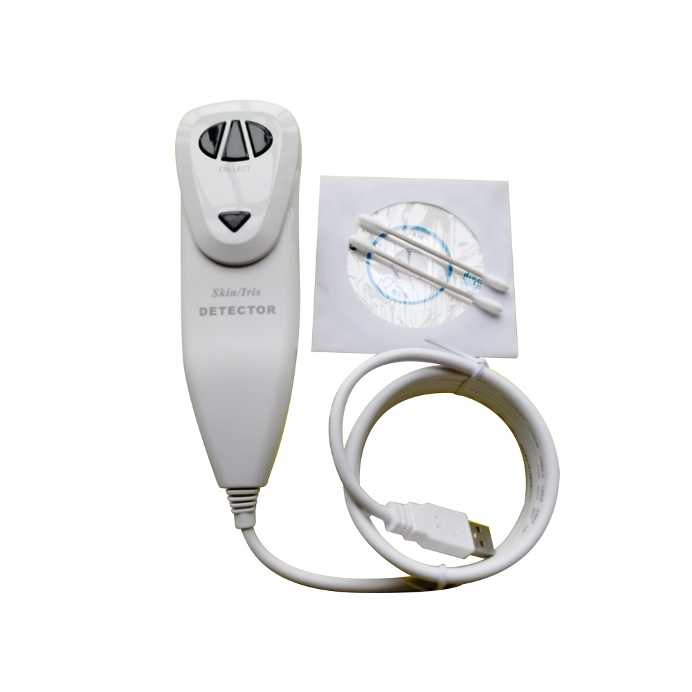
The human cornea functions as a curved, wet mirror. When direct light hits it, a harsh reflection bounces back into the lens, obscuring the pupil and surrounding tissue. Standard ring lights often create a circular flash that obliterates the collarette.
A specialized Iris Scanner solves this dilemma using deliberate lighting geometries. Engineers set LEDs at precise oblique angles or use polarized side-illumination channels. This architecture directs reflections away from the central optical path, revealing fine structural fibers across the iris face.
Practitioners should observe the following lighting guidelines during examinations:
Use bilateral light sources to illuminate the nasal and temporal quadrants evenly.
Avoid unshielded overhead fluorescent or direct sunlight in the evaluation room.
Employ angled single-side lighting when evaluating deep stromal depressions and crypt relief.
Check that the corneal reflex falls safely outside the autonomic nerve ring area.
Inexpensive LED diodes often emit discontinuous light spectra with sharp blue peaks. This artificial cast ruins color balance and causes pupil constriction during exams. Professional equipment utilizes high Color Rendering Index (CRI ≥ 95) LEDs balanced between 5000K and 5500K.
This daylight-equivalent spectrum reveals true pigment depth. It prevents blue eyes from appearing steel-gray and protects brown eyes from muddy, unreadable visual tones. The balanced wavelength distribution illuminates the tissue without generating heat or causing client discomfort.
No two eyes absorb light the same way. A dense, dark brown iris requires strong light penetration to highlight hidden crypts. In contrast, a light blue or albino-variant iris easily overexposes under moderate light. Modern devices include multi-step hardware dimmers, giving you full control over light intensity for every client profile.
Standard cameras rely on digital zoom, cropping an image and interpolating pixels. This approach introduces blur and ruins detail. In contrast, a clinical-grade Iridology Camera uses dedicated optical glass lenses engineered for true macro magnification. These lenses minimize optical aberrations and maintain razor-sharp clarity from edge to edge.
Autofocus systems struggle with ocular anatomy. Most standard autofocus algorithms hunt for contrast, locking onto eyelashes, tear film bubbles, or eyelid margins instead of the iris plane. This hunting frustrates practitioners and agitates the client's eye.
Clinical practice demands smooth, manual micro-focus control. A tactile focus wheel allows practitioners to guide the focal plane directly onto the trabecular meshwork. Some hybrid systems offer continuous focus assist, but manual override remains essential for pinpoint adjustments.
Feature | Entry-Level / Generic Scope | Clinical Macro Iridology Camera |
|---|---|---|
Lens Type | Single-layer acrylic element | Multi-coated optical glass macro lens |
Focus Control | Fixed focus or coarse autofocus | Smooth manual micro-adjustment dial |
Illumination Geometry | Direct front-facing ring light | Dual-angle, glare-deflecting cold LED array |
Color Rendering (CRI) | CRI < 75 (High blue/green drift) | CRI ≥ 95 (Full spectrum daylight 5000–5500K) |
Distortion Profile | Noticeable barrel distortion at edges | Flat-field optical correction |
The iris is not flat. It curves outward across the anterior chamber, featuring structural valleys and raised collarettes. A narrow depth of field leaves the pupil margin sharp while blurring the outer ciliary zone. Clinical systems balance focal length with an optimized fixed aperture (typically f/8 to f/11), keeping the entire ocular surface sharp in a single shot.
High-end hardware needs capable software to complete the diagnostic workflow. Once an image is captured, software takes over to organize, analyze, and store the clinical file safely. Modern clinics cannot afford cumbersome file structures or disconnected storage setups.
High-resolution iris imagery qualifies as biometric data under privacy frameworks like HIPAA in the United States and GDPR in the European Union. You must manage these visual files with the same diligence as electronic medical records. Your software platform must provide local data encryption, password authentication, and safe backup systems.
Practitioners should watch out for cheap devices that rely on unsecured, third-party cloud apps. Storing unencrypted eye files on foreign servers can expose clinics to serious compliance penalties. Prioritize systems that support local storage on your clinic workstation or secure, HIPAA-compliant networks.
Modern software suites improve consultation efficiency with practical clinical tools:
Transparent Bernard Jensen, Rayid, or Angerer chart overlays scaled automatically to the pupil-iris ratio.
Side-by-side split screens comparing historical scans to highlight subtle tissue shifts over time.
Quadrant-based digital magnifying loupes for isolated review of micro-signs.
Neutral contrast and greyscale toggles to examine fine scleral vessels without color distractions.
Be cautious with vendors promoting "automated AI diagnosis." These programs claim to detect systemic diseases from an iris photo. Such claims lack clinical validation and expose practices to medical liability risks. The best software aids documentation, measurement, and workflow without making speculative diagnostic claims.
Selecting equipment involves assessing your clinic's physical environment and consultation flow. A scanner with exceptional optics will fail your practice if it feels awkward to hold or crashes your computer. Focus on practical mechanics and usability when making your choice.
Iridology devices generally come in three forms: handheld wands, articulated boom arms, and slit-lamp chin-rest stations. Each design suits different clinical spaces:
Handheld Wands: Lightweight and portable, ideal for traveling practitioners or multi-room clinics. Look for steady grips and built-in finger triggers.
Boom Arm Mounts: These balance the camera on a counterweight arm, eliminating camera shake while keeping the client comfortable in their chair.
Chin-Rest Tabletop Units: The gold standard for stability. They immobilize the client's head, guaranteeing repeatable distances and sharp focal planes.
Ensure your chosen camera uses standard USB Video Class (UVC) protocols. Proprietary drivers often break when operating systems update. Verify whether the software works smoothly across both Windows and macOS platforms, and ask the manufacturer about their history of software updates.
Before purchasing, follow these recommended evaluation steps:
Request uncompressed, raw image samples from both light and dark eyes.
Schedule a live software demonstration to inspect file management and overlay adjustments.
Review warranty terms and clarify replacement turnaround times to protect against downtime.
Confirm that the hardware runs directly through powered USB ports without requiring cumbersome external power supplies.
Selecting a professional iridology camera requires balancing optical clarity, balanced illumination, manual focus control, and secure software. By investing in dedicated equipment rather than consumer gadgets, you protect assessment accuracy and elevate client trust. High-fidelity imaging turns visual evaluations into reliable, repeatable clinical records.
Audit your current imaging setup today. Check your lighting for color accuracy, test your focus sharpness at deep zoom levels, and ensure your software complies with patient privacy standards. Reach out to specialized optical manufacturers, request raw image samples, and select equipment that serves your practice for years to come.
A: An 8MP iridology camera is the professional benchmark. It provides sharp pixel density for deep magnification into iris crypts and trabeculae without overwhelming your computer storage with oversized files.
A: This reflection is corneal glare from direct, unshielded light. Professional iris systems use angled, dual-source, or polarized illumination engineered to bounce reflections away from the central optical lens.
A: Smartphones rely on aggressive software processing, artificial sharpening, and dynamic tone adjustments. These automated changes alter real colors and hide subtle tissue structures, making them unsuitable for reliable clinical evaluations.
A: While the camera hardware can capture raw photos on its own, dedicated software is essential. It provides secure client record management, interactive chart overlays, and side-by-side historical comparisons for ongoing care.


